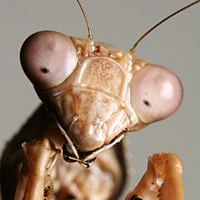
In the compound eye of invertebrates such as insects and crustaceans, the pseudopupil appears as a dark spot which moves across the eye as the animal is rotated.[1] This occurs because the ommatidia that one observes "head-on" (along their optical axes) absorb the incident light, while those to one side reflect it.[2] The pseudopupil therefore reveals which ommatidia are aligned with the axis along which the observer is viewing.[2]


Pseudopupil analysis technique
editThe pseudopupil analysis technique is used to study neurodegeneration in insects like Drosophila. An adult Drosophila eye consists of nearly 800 unit ommatidia which are repeated in a symmetrical pattern. Each ommatidium contains 8 photoreceptor cells, each of which forms a rhabdomere (rhabdomeres 7 and 8 overlap vertically; therefore, only rhabdomere 7 is visible externally). Neurodegeneration leads to loss or degradation of photoreceptors.[3] By visualizing and counting the intact rhabdomeres, degradation level can be measured. Thus, analyzing the pseudopupil can permit empirical study of neurodegeneration.
References
edit- ^ M. F. Land; G. Gibson; J. Horwood; J. Zeil (1999). "Fundamental differences in the optical structure of the eyes of nocturnal and diurnal mosquitoes" (PDF). Journal of Comparative Physiology A. 185 (1): 91–103. doi:10.1007/s003590050369. S2CID 9114187. Archived from the original (PDF) on 2016-03-04. Retrieved 2008-07-27.
- ^ a b Jochen Zeil & Maha M. Al-Mutairi (1996). "Variations in the optical properties of the compound eyes of Uca lactea annulipes" (PDF). The Journal of Experimental Biology. 199 (7): 1569–1577. PMID 9319471.
- ^ Song, Wan; Smith, Marianne R.; Syed, Adeela; Lukacsovich, Tamas; Barbaro, Brett A.; Purcell, Judith; Bornemann, Doug J.; Burke, John; Marsh, J. Lawrence (2013). Morphometric analysis of Huntington's disease neurodegeneration in Drosophila. Methods in Molecular Biology. Vol. 1017. pp. 41–57. doi:10.1007/978-1-62703-438-8_3. ISBN 978-1-62703-437-1. ISSN 1940-6029. PMID 23719906.