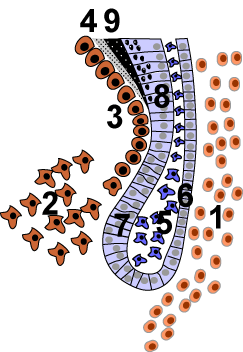
In animal tooth development, the inner enamel epithelium, also known as the internal enamel epithelium, is a layer of columnar cells located on the rim nearest the dental papilla of the enamel organ in a developing tooth. This layer is first seen during the cap stage, in which these inner enamel epithelium cells are pre-ameloblast cells. These will differentiate into ameloblasts which are responsible for secretion of enamel during tooth development.
| Inner enamel epithelium | |
|---|---|
 The cervical loop area: (1) dental follicle cells, (2) dental mesenchyme, (3) odontoblasts, (4) dentin, (5) stellate reticulum, (6) outer enamel epithelium, (7) inner enamel epithelium, (8) ameloblasts, (9) enamel. | |
| Details | |
| Identifiers | |
| Latin | epithelium enameleum internum |
| TE | enamel epithelium_by_E5.4.1.1.2.3.15 E5.4.1.1.2.3.15 |
| Anatomical terminology | |
The location of the enamel organ where the outer and inner enamel epithelium join is called the cervical loop.
References
edit- Cate, A.R. Ten. Oral Histology: development, structure, and function. 5th ed. 1998. ISBN 0-8151-2952-1.
- Ross, Michael H., Gordon I. Kaye, and Wojciech Pawlina. Histology: a text and atlas. 4th edition. 2003. ISBN 0-683-30242-6.