In molecular biology, Glycoside hydrolase family 2 is a family of glycoside hydrolases EC 3.2.1., which are a widespread group of enzymes that hydrolyse the glycosidic bond between two or more carbohydrates, or between a carbohydrate and a non-carbohydrate moiety. A classification system for glycoside hydrolases, based on sequence similarity, has led to the definition of >100 different families.[1][2][3] This classification is available on the CAZy web site,[4][5] and also discussed at CAZypedia, an online encyclopedia of carbohydrate active enzymes.[6][7]
| Glycosyl hydrolases family 2, sugar binding domain | |||||||||
|---|---|---|---|---|---|---|---|---|---|
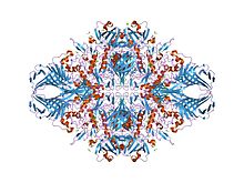
 e. coli (lacz) beta-galactosidase-trapped 2-deoxy-galactosyl enzyme intermediate | |||||||||
| Identifiers | |||||||||
| Symbol | Glyco_hydro_2_N | ||||||||
| Pfam | PF02837 | ||||||||
| Pfam clan | CL0202 | ||||||||
| InterPro | IPR006104 | ||||||||
| PROSITE | PDOC00531 | ||||||||
| SCOP2 | 1bgl / SCOPe / SUPFAM | ||||||||
| CAZy | GH2 | ||||||||
| |||||||||
| Glycosyl hydrolases family 2 | |||||||||
|---|---|---|---|---|---|---|---|---|---|
 e. coli (lacz) beta-galactosidase-trapped 2-deoxy-galactosyl enzyme intermediate | |||||||||
| Identifiers | |||||||||
| Symbol | Glyco_hydro_2 | ||||||||
| Pfam | PF00703 | ||||||||
| InterPro | IPR006102 | ||||||||
| PROSITE | PDOC00531 | ||||||||
| SCOP2 | 1bgl / SCOPe / SUPFAM | ||||||||
| CAZy | GH2 | ||||||||
| |||||||||
| Glycosyl hydrolases family 2, TIM barrel domain | |||||||||
|---|---|---|---|---|---|---|---|---|---|
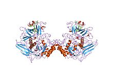
 human beta-glucuronidase at 2.6 a resolution | |||||||||
| Identifiers | |||||||||
| Symbol | Glyco_hydro_2_C | ||||||||
| Pfam | PF02836 | ||||||||
| Pfam clan | CL0058 | ||||||||
| InterPro | IPR006103 | ||||||||
| PROSITE | PDOC00531 | ||||||||
| SCOP2 | 1bgl / SCOPe / SUPFAM | ||||||||
| CAZy | GH2 | ||||||||
| |||||||||
Glycoside hydrolase family 2[8] comprises enzymes with several known activities: beta-galactosidase (EC 3.2.1.23); beta-mannosidase (EC 3.2.1.25); beta-glucuronidase (EC 3.2.1.31). These enzymes contain a conserved glutamic acid residue which has been shown,[9] in Escherichia coli lacZ (P00722), to be the general acid/base catalyst in the active site of the enzyme.
The catalytic domain of Beta-galactosidases have a TIM barrel core surrounded several other largely beta domains.[10] The sugar binding domain of these proteins has a jelly-roll fold.[10] These enzymes also include an immunoglobulin-like beta-sandwich domain.[10]
External links
editReferences
edit- ^ Henrissat B, Callebaut I, Fabrega S, Lehn P, Mornon JP, Davies G (July 1995). "Conserved catalytic machinery and the prediction of a common fold for several families of glycosyl hydrolases". Proceedings of the National Academy of Sciences of the United States of America. 92 (15): 7090–4. Bibcode:1995PNAS...92.7090H. doi:10.1073/pnas.92.15.7090. PMC 41477. PMID 7624375.
- ^ Davies G, Henrissat B (September 1995). "Structures and mechanisms of glycosyl hydrolases". Structure. 3 (9): 853–9. doi:10.1016/S0969-2126(01)00220-9. PMID 8535779.
- ^ Henrissat B, Bairoch A (June 1996). "Updating the sequence-based classification of glycosyl hydrolases". The Biochemical Journal. 316 (Pt 2): 695–6. doi:10.1042/bj3160695. PMC 1217404. PMID 8687420.
- ^ "Home". CAZy.org. Retrieved 2018-03-06.
- ^ Lombard V, Golaconda Ramulu H, Drula E, Coutinho PM, Henrissat B (January 2014). "The carbohydrate-active enzymes database (CAZy) in 2013". Nucleic Acids Research. 42 (Database issue): D490-5. doi:10.1093/nar/gkt1178. PMC 3965031. PMID 24270786.
- ^ "Glycoside Hydrolase Family 2". CAZypedia.org. Retrieved 2018-03-06.
- ^ CAZypedia Consortium (December 2018). "Ten years of CAZypedia: a living encyclopedia of carbohydrate-active enzymes" (PDF). Glycobiology. 28 (1): 3–8. doi:10.1093/glycob/cwx089. PMID 29040563.
- ^ Withers S. "Glycoside Hydrolase Family 2 (GH_2)". CAZypedia - carbohydrate active enzymes. Retrieved 2010-10-10.
- ^ Gebler JC, Aebersold R, Withers SG (June 1992). "Glu-537, not Glu-461, is the nucleophile in the active site of (lac Z) beta-galactosidase from Escherichia coli". The Journal of Biological Chemistry. 267 (16): 11126–30. doi:10.1016/S0021-9258(19)49884-0. PMID 1350782.
- ^ a b c Jacobson RH, Zhang XJ, DuBose RF, Matthews BW (June 1994). "Three-dimensional structure of beta-galactosidase from E. coli". Nature. 369 (6483): 761–6. Bibcode:1994Natur.369..761J. doi:10.1038/369761a0. PMID 8008071. S2CID 4241867.