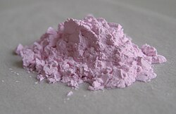
Erbium(III) oxide is the inorganic compound with the formula Er2O3. It is a pink paramagnetic solid. It finds uses in various optical materials.[2]

| |

| |
| Names | |
|---|---|
| Other names
Erbium oxide, erbia
| |
| Identifiers | |
3D model (JSmol)
|
|
| ChemSpider | |
| ECHA InfoCard | 100.031.847 |
| EC Number |
|
PubChem CID
|
|
CompTox Dashboard (EPA)
|
|
| |
| |
| Properties | |
| Er2O3 | |
| Molar mass | 382.56 g/mol |
| Appearance | pink crystals |
| Density | 8.64 g/cm3 |
| Melting point | 2,344 °C (4,251 °F; 2,617 K) |
| Boiling point | 3,290 °C (5,950 °F; 3,560 K) |
| insoluble in water | |
| +73,920·10−6 cm3/mol | |
| Structure | |
| Cubic, cI80 | |
| Ia-3, No. 206 | |
| Thermochemistry | |
Heat capacity (C)
|
108.5 J·mol−1·K−1 |
Std molar
entropy (S⦵298) |
155.6 J·mol−1·K−1 |
Std enthalpy of
formation (ΔfH⦵298) |
−1897.9 kJ·mol−1 |
| Related compounds | |
Other anions
|
Erbium(III) chloride |
Other cations
|
Holmium(III) oxide, Thulium(III) oxide |
Except where otherwise noted, data are given for materials in their standard state (at 25 °C [77 °F], 100 kPa).
| |
Structure
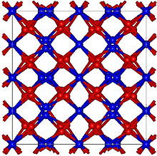
editErbium(III) oxide has a cubic structure resembling the bixbyite motif. The Er3+ centers are octahedral.[2]
Reactions
editErbium oxide is produced by burning erbium metal.[3] Erbium oxide is insoluble in water but soluble in mineral acids. Er2O3 does not readily absorb moisture and carbon dioxide from the atmosphere. It can react with acids to form the corresponding erbium(III) salts. For example, with hydrochloric acid, the oxide follows the following idealized reaction leading to erbium chloride:
- Er2O3 + 6 HCl → 2 ErCl3 + 3 H2O
In practice, such simple acid-base reactions are accompanied by hydration:
- ErCl3 + 9 H2O → [Er(H2O)9]Cl3
Properties
editOne interesting property of erbium oxides is their ability to up convert photons. Photon upconversion takes place when infrared or visible radiation, low energy light, is converted to ultraviolet or violet radiation higher energy light via multiple transfer or absorption of energy.[4] Erbium oxide nanoparticles also possess photoluminescence properties. Erbium oxide nanoparticles can be formed by applying ultrasound (20 kHz, 29 W·cm−2) in the presence of multiwall carbon nanotubes. The erbium oxide nanoparticles that have been produced using ultrasound are erbium carboxioxide, hexagonal and spherical geometry erbium oxide. Each ultrasonically formed erbium oxide exhibits photoluminescence in the visible region of the electromagnetic spectrum under excitation of wavelength 379 nm in water. Hexagonal erbium oxide photoluminescence is long-lived and allows higher energy transitions (4S3/2 – 4I15/2). Spherical erbium oxide does not undergo 4S3/2 – 4I15/2 energy transitions.[5]
Uses
editThe applications of Er2O3 are varied due to their electrical, optical and photoluminescence properties. Nanoscale materials doped with Er3+ are of much interest because they have special particle-size-dependent optical and electrical properties.[6] Erbium oxide doped nanoparticle materials can be dispersed in glass or plastic for display purposes, such as display monitors. The spectroscopy of Er3+ electronic transitions in host crystals lattices[clarification needed][words missing?] of nanoparticles combined with ultrasonically formed geometries in aqueous solution of carbon nanotubes is of great interest for synthesis of photoluminescence nanoparticles in "green" chemistry.[5]
Erbium oxide is widely used in interferometers that require high-power lasers.[7] These interferometers often employ erbium-doped fiber amplifiers (EDFAs) to enhance the power of the laser beams.[8] EDFAs, which utilize erbium ions, provide low noise and high gain, making them ideal for long-distance signal transmission and high-resolution measurements in interferometry.[9]
Erbium oxide is among the most important rare earth metals used in biomedicine.[10] The photoluminescence property of erbium oxide nanoparticles on carbon nanotubes makes them useful in biomedical applications. For example, erbium oxide nanoparticles can be surface modified for distribution into aqueous and non-aqueous media for bioimaging.[6] Erbium oxides are also used as gate dielectrics in semiconductor devices since it has a high dielectric constant (10–14) and a large band gap. Erbium is sometimes used as a coloring for glasses,[1] and erbium oxide can also be used as a burnable neutron poison for nuclear fuel.
History
editImpure erbium(III) oxide was isolated by Carl Gustaf Mosander in 1843, and first obtained in pure form in 1905 by Georges Urbain and Charles James.[11]
References
edit- ^ a b Lide, David R. (1998). Handbook of Chemistry and Physics (87 ed.). Boca Raton, FL: CRC Press. pp. 4–57. ISBN 978-0-8493-0594-8.
- ^ a b Adachi, Gin-ya; Imanaka, Nobuhito (1998). "The Binary Rare Earth Oxides". Chemical Reviews. 98 (4): 1479–1514. doi:10.1021/cr940055h. PMID 11848940.
- ^ Emsley, John (2001). "Erbium" Nature's Building Blocks: An A-Z Guide to Elements. Oxford, England, Uk: Oxford University Press. pp. 136–139. ISBN 978-0-19-850340-8.
- ^ "Rare-earth-doped nanoparticles prove illuminating". SPIE. Retrieved April 10, 2012.
- ^ a b Radziuk, Darya; André Skirtach; Andre Geßner; Michael U. Kumke; Wei Zhang; Helmuth Möhwald; Dmitry Shchukin (24 October 2011). "Ultrasonic Approach for Formation of Erbium Oxide Nanoparticles with Variable Geometries". Langmuir. 27 (23): 14472–14480. doi:10.1021/la203622u. PMID 22022886.
- ^ a b Richard, Scheps (12 February 1996). "Upconversion laser processes". Progress in Quantum Electronics. 20 (4): 271–358. Bibcode:1996PQE....20..271S. doi:10.1016/0079-6727(95)00007-0.
- ^ Li, Chunfei; Wang, Fei (2007). "Optimization of all-optical EDFA-based Sagnac-interferometer switch". Optics Express. 15 (21): 14234–14243. Bibcode:2007OExpr..1514234W. doi:10.1364/OE.15.014234. PMID 19550698.
- ^ Lawen, Eric. "Applications of Erbium Oxide in Glass Production". Stanford Advanced Materials. Retrieved July 26, 2024.
- ^ Kaler, Rajneesh; Kaler, R.S. (2011). "Gain and Noise figure performance of erbium doped fiber amplifiers (EDFAs) and Compact EDFAs". Optik. 122 (5): 440–443. Bibcode:2011Optik.122..440K. doi:10.1016/j.ijleo.2010.02.028.
- ^ Andre, Skirtach; Almudena Javier; Oliver Kref; Karen Kohler; Alicia Alberola; Helmuth Mohwald; Wolfgang Parak; Gleb Sukhorukov (2006). "Laser-Induced Release of Encapsulated Materials inside Living Cells" (PDF). Angew. Chem. Int. Ed. 38 (28): 4612–4617. doi:10.1002/anie.200504599. PMID 16791887. Retrieved April 15, 2012.
- ^ Aaron John Ihde (1984). The development of modern chemistry. Courier Dover Publications. pp. 378–379. ISBN 978-0-486-64235-2.