Ectopia lentis is a displacement or malposition of the eye's lens from its normal location. A partial dislocation of a lens is termed lens subluxation or subluxated lens; a complete dislocation of a lens is termed lens luxation or luxated lens.
| Ectopia lentis | |
|---|---|
 | |
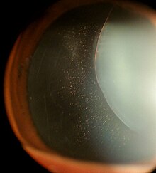
| Ectopia Lentis in Marfan syndrome. Zonular fibers are being seen. | |
| Specialty | Medical genetics |


Ectopia lentis in dogs and cats
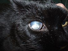
editAlthough observed in humans and cats, ectopia lentis is most commonly seen in dogs. Ciliary zonules normally hold the lens in place. Abnormal development of these zonules can lead to primary ectopia lentis, usually a bilateral condition. Luxation can also be a secondary condition, caused by trauma, cataract formation (decrease in lens diameter may stretch and break the zonules), or glaucoma (enlargement of the globe stretches the zonules). Steroid administration weakens the zonules and can lead to luxation, as well. Lens luxation in cats can occur secondary to anterior uveitis (inflammation of the inside of the eye).
Anterior lens luxation
editWith anterior lens luxation, the lens pushes into the iris or actually enters the anterior chamber of the eye. This can cause glaucoma, uveitis, or damage to the cornea. Uveitis (inflammation of the eye) causes the pupil to constrict (miosis) and trap the lens in the anterior chamber, leading to an obstruction of outflow of aqueous humour and subsequent increase in ocular pressure (glaucoma).[1] Better prognosis is valued in lens replacement surgery (retained vision and normal intraocular pressure) when it is performed before the onset of secondary glaucoma.[2] Glaucoma secondary to anterior lens luxation is less common in cats than dogs due to their naturally deeper anterior chamber and the liquification of the vitreous humour secondary to chronic inflammation.[3] Anterior lens luxation is considered to be an ophthalmological emergency.
Posterior lens luxation
editWith posterior lens luxation, the lens falls back into the vitreous humour and lies on the floor of the eye. This type causes fewer problems than anterior lens luxation, although glaucoma or ocular inflammation may occur. Surgery is used to treat dogs with significant symptoms. Removal of the lens before it moves to the anterior chamber may prevent secondary glaucoma.[2]
Lens subluxation
editLens subluxation is also seen in dogs and is characterized by a partial displacement of the lens. It can be recognized by trembling of the iris (iridodonesis) or lens (phacodonesis) and the presence of an aphakic crescent (an area of the pupil where the lens is absent).[4] Other signs of lens subluxation include mild conjunctival redness, vitreous humour degeneration, prolapse of the vitreous into the anterior chamber, and an increase or decrease of anterior chamber depth.[5] Removal of the lens before it completely luxates into the anterior chamber may prevent secondary glaucoma.[2] Extreme degree of luxation of lens is called "lenticele" in which lens comes out of the eyeball and becomes trapped under the Tenon's capsule or conjunctiva.[6] A nonsurgical alternative treatment involves the use of a miotic to constrict the pupil and prevent the lens from luxating into the anterior chamber.[7]
Breed predisposition
editTerrier breeds are predisposed to lens luxation, and it is probably inherited in the Sealyham Terrier, Jack Russell Terrier, Wirehaired Fox Terrier, Rat Terrier, Teddy Roosevelt Terrier, Tibetan Terrier,[8] Miniature Bull Terrier, Shar Pei, and Border Collie.[9] The mode of inheritance in the Tibetan Terrier[5] and Shar Pei[10] is likely autosomal recessive. Labrador Retrievers and Australian Cattle Dogs are also predisposed.[11]
Systemic associations in humans
editIn humans, a number of systemic conditions are associated with ectopia lentis:[12]
More common:
- Marfan syndrome (upward and outward)[13]
- Homocystinuria (downward and inwards)[13]
- Weill–Marchesani syndrome
- Sulfite oxidase deficiency
- Molybdenum cofactor deficiency
- Hyperlysinemia
Less common:
See also
editReferences
edit- ^ Ketring, Kerry I. (2006). "Emergency Treatment for Anterior Lens Luxation" (PDF). Proceedings of the North American Veterinary Conference. Archived from the original (PDF) on 2007-09-29. Retrieved 2007-02-22.
- ^ a b c Glover T, Davidson M, Nasisse M, Olivero D (1995). "The intracapsular extraction of displaced lenses in dogs: a retrospective study of 57 cases (1984-1990)". Journal of the American Animal Hospital Association. 31 (1): 77–81. doi:10.5326/15473317-31-1-77. PMID 7820769.
- ^ Peiffer, Robert L. Jr. (2004). "Diseases of the Lens in Dogs and Cats". Proceedings of the 29th World Congress of the World Small Animal Veterinary Association. Retrieved 2007-02-22.
- ^ "Lens". The Merck Veterinary Manual. 2006. Retrieved 2007-02-22.
- ^ a b Grahn B, Storey E, Cullen C (2003). "Diagnostic ophthalmology. Congenital lens luxation and secondary glaucoma". Canadian Veterinary Journal. 44 (5): 427, 429–30. PMC 340155. PMID 12757137.
- ^ Shah SIA et al: Concise Ophthalmology Text & Atals. 5th ed. Param B (Pvt.) Ltd. 2018: 60-61
- ^ Binder DR, Herring IP, Gerhard T (2007). "Outcomes of nonsurgical management and efficacy of demecarium bromide treatment for primary lens instability in dogs: 34 cases (1990-2004)". Journal of the American Veterinary Medical Association. 231 (1): 89–93. doi:10.2460/javma.231.1.89. PMID 17605669.
- ^ Gelatt, Kirk N., ed. (1999). Veterinary Ophthalmology (3rd ed.). Lippincott, Williams & Wilkins. ISBN 0-683-30076-8.
- ^ Petersen-Jones, Simon M. (2003). "Conditions of the Lens". Proceedings of the 28th World Congress of the World Small Animal Veterinary Association. Retrieved 2007-02-22.
- ^ Lazarus J, Pickett J, Champagne E (1998). "Primary lens luxation in the Chinese Shar Pei: clinical and hereditary characteristics". Veterinary Ophthalmology. 1 (2–3): 101–107. doi:10.1046/j.1463-5224.1998.00021.x. PMID 11397217.
- ^ Johnsen D, Maggs D, Kass P (2006). "Evaluation of risk factors for development of secondary glaucoma in dogs: 156 cases (1999-2004)". Journal of the American Veterinary Medical Association. 229 (8): 1270–4. doi:10.2460/javma.229.8.1270. PMID 17042730.
- ^ Eifrig CW, Eifrig DE. "Ectopia Lentis". eMedicine.com. November 24, 2004.
- ^ a b Peter Nicholas Robinson; Maurice Godfrey (2004). Marfan syndrome: a primer for clinicians and scientists. Springer. pp. 5–. ISBN 978-0-306-48238-0. Retrieved 12 April 2010.