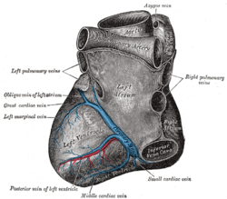
The smallest cardiac veins (also known as the Thebesian veins (named for Adam Christian Thebesius)) are small, valveless veins in the walls of all four heart chambers[1] that drain venous blood from the myocardium[2] directly into any of the heart chambers.[3]
| Smallest cardiac veins | |
|---|---|
 | |
| Details | |
| Identifiers | |
| Latin | venae cardiacae minimae, venae cordis minimae |
| TA98 | A12.3.01.013 |
| TA2 | 4169 |
| FMA | 71568 |
| Anatomical terminology | |
They are most abundant in the right atrium, and least abundant in the left ventricle.[4][better source needed]
Structure
editThe smallest cardiac veins vary greatly in size and number. Those draining the right atrium have a lumen of up to 2 mm in diameter, whereas those draining the right ventricle have lumens as small as 0.5 mm in diameter.[5]
Course
editThey run a perpendicular course to the endocardial surface, directly connecting the heart chambers to the medium-sized, and larger coronary veins.[6]
Openings
editThe openings of the smallest cardiac veins are located in the endocardium. Here the smallest cardiac veins return blood into the heart chambers from the capillary bed in the muscular cardiac wall, enabling a form of collateral circulation unique to the heart. Not every endocardial opening connects to the smallest cardiac veins, as some connect to the vessels of Wearn, which are arteries. Therefore, the endocardial opening must be traced to a vein before it is definitely called an opening of the smallest cardiac veins.[citation needed]
Function
editThe small cardiac veins are responsible for venous return of 10% of the coronary blood supply.[7] The small cardiac venous network is considered an alternative venous drainage of the myocardium. The smallest cardiac veins draining into the left heart, along with deoxygenated blood originating from the bronchial veins draining into the pulmonary veins, contribute to normal physiologic shunting of blood. As a consequence of the input of these vessels, blood in the left heart is less oxygenated than the blood found at the pulmonary capillary beds, usually to a very small degree.
History
editEtymology
editThe smallest cardiac veins are also known as the Thebesian veins.[8][9] They are named after the German anatomist Adam Christian Thebesius, who described them in a 1708 treatise called Disputatio medica inauguralis de circulo sanguinis in corde.[10][11]
Mislabelling
editThe smallest cardiac veins are sometimes accurately referred to as vessels, but they are frequently confused with a distinct set of artery connections,[12][13][14][15] eponymously referred to as the "vessels of Wearn".[16] In his 1928 publication, Wearn himself referred to the arterio-cameral connections (vessels of Wearn) as Thebesian,[17] but later, after additional research, provided disambiguation and strictly used the term Thebesian for coronary vein-heart chamber connections.[18]
References
edit- ^ Blake, HA; Manion, WC; Mattingly, TW; Baroldi, G (1964). "Coronary artery anomalies". Circulation. 30 (6): 927–40. doi:10.1161/01.cir.30.6.927. PMID 14246341.
- ^ Agur, AMR; Dalley, AF (2009). Grant's atlas of anatomy. Lippincott Williams & Wilkins. pp. 53–. ISBN 978-0-7817-7055-2. Retrieved 31 October 2010.
- ^ Wilson, Alexander; Bhutta, Beenish S. (2022), "Anatomy, Thorax, Coronary Sinus", StatPearls, Treasure Island (FL): StatPearls Publishing, PMID 32491498, retrieved 2023-01-05
- ^ "Venae cordis minimae". radiopaedia.org. Retrieved 31 August 2019.
- ^ Standring, Susan (2020). Gray's Anatomy: The Anatomical Basis of Clinical Practice (42th ed.). New York. p. 1093. ISBN 978-0-7020-7707-4. OCLC 1201341621.
{{cite book}}: CS1 maint: location missing publisher (link) - ^ Pratt, FH (1898). "The nutrition of the heart through the vessels of Thebesius and the cardiac veins" (PDF). American Journal of Physiology. 1: 86–103. doi:10.1152/ajplegacy.1898.1.1.86.
- ^ Wilson, Alexander; Bhutta, Beenish S. (2022), "Anatomy, Thorax, Coronary Sinus", StatPearls, Treasure Island (FL): StatPearls Publishing, PMID 32491498, retrieved 2023-01-05
- ^ Singhal, S; Khoury, S (2008). "Images in clinical medicine. Imaging of thebesian venous system". The New England Journal of Medicine. 359 (7): e8. doi:10.1056/nejmicm072885. PMID 18703466.
- ^ Smith, TM (2009). "Rare view of thebesian venous system". Radiologic Technology. 81 (2): 173–4. PMID 19901354.
- ^ synd/4013 at Who Named It?
- ^ Thebesius, AC (1708). Disputatio medica inauguralis de circulo sanguinis in corde. Doctoral dissertation, Leiden.
- ^ Boeder, NF; Nef, HM; Bauer, T (2016). "Thebesian veins as drainage to the ventricle: A case report". Cardiovascular Revascularization Medicine. 18 (3): 213–214. doi:10.1016/j.carrev.2016.09.007. PMID 27743821.
- ^ Grollman, JH Jr (1998). "Re: three major coronary artery-to-left ventricular shunts". CardioVascular and Interventional Radiology. 21 (2): 183. doi:10.1007/s002709900240. PMID 9502691. S2CID 40212007.
- ^ Wearn, JT (1941). "Morphological and functional alterations of the coronary circulation". Bulletin of the New York Academy of Medicine. 17 (10): 754–777. PMC 1933738. PMID 19312228.
- ^ Wearn, JT; Mettier, SR; Klumpp, TG; Zschiesche, LJ (1933). "The nature of the vascular communications between the coronary arteries and the chambers of the heart". American Heart Journal. 9 (2): 143–164. doi:10.1016/S0002-8703(33)90711-5.
- ^ Hussain, M; Roberts, EB (2015). "Association of coronary to left ventricular microfistulae (vessels of Wearn) with atrial septal defect in an adult without cyanotic heart disease". BMJ Case Reports. 2015: bcr2014207655. doi:10.1136/bcr-2014-207655. PMC 4493239. PMID 26139649.
- ^ Wearn, JT (1928). "The role of the thebesian vessels in the circulation of the heart". Journal of Experimental Medicine. 47 (2): 293–315. doi:10.1084/jem.47.2.293. PMC 2131354. PMID 19869414.
- ^ Wearn, JT; Mettier, SR; Klumpp, TG; Zschiesche, LJ (1933). "The nature of the vascular communications between the coronary arteries and the chambers of the heart". American Heart Journal. 9 (2): 143–164. doi:10.1016/S0002-8703(33)90711-5.