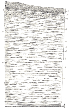
Descemet's membrane (or the Descemet membrane) is the basement membrane that lies between the corneal proper substance, also called stroma, and the endothelial layer of the cornea. It is composed of different kinds of collagen (Type IV and VIII)[1] than the stroma. The endothelial layer is located at the posterior of the cornea. Descemet's membrane, as the basement membrane for the endothelial layer, is secreted by the single layer of squamous epithelial cells that compose the endothelial layer of the cornea.
| Descemet's membrane | |
|---|---|
 Vertical section of human cornea from near the margin. (Waldeyer.) Magnified.
| |
| Details | |
| Pronunciation | English: /ˈdɛsəmeɪ/ |
| Location | Cornea of eye |
| Identifiers | |
| Latin | l. limitans posterior corneae |
| MeSH | D003886 |
| TA98 | A15.2.02.021 |
| FMA | 58309 |
| Anatomical terms of microanatomy | |
Structure
editIts thickness ranges from 3 μm at birth to 8–10 μm in adults.[2]
The corneal endothelium is a single layer of squamous cells covering the surface of the cornea that faces the anterior chamber.
Clinical significance
editSignificant damage to the membrane may require a corneal transplant. Damage caused by the hereditary condition known as Fuchs dystrophy (q.v.)—where Descemet's membrane progressively fails and the cornea thickens and clouds because the exchange of nutrients/fluids between the cornea and the rest of the eye is interrupted—can be reversed by surgery. The surgeon can scrape away the damaged Descemet membrane and insert/transplant a new membrane harvested from the eye of a donor.[3] In the process most of the squamous cells of the donor membrane survive to dramatically and emphatically reverse the corneal deterioration (see DMEK surgery).
Descemet's membrane is also a site of copper deposition in patients with Wilson's disease or other liver diseases, leading to formation of Kayser–Fleischer rings.
History
editIt is also known as the posterior limiting elastic lamina, lamina elastica posterior, and membrane of Demours. It was named after French physician Jean Descemet (1732–1810).
See also
editReferences
edit- ^ "Tissue Distribution of Type VIII Collagen in Human Adult and Fetal Eyes" (PDF). Investigative Ophthalmology and Visual Science. 1991-08-01. Retrieved 2014-08-17.
- ^ Johnson DH, Bourne WM, Campbell RJ: The ultrastructure of Descemet's membrane. I. Changes with age in normal cornea. Arch Ophthalmol 100:1942, 1982
- ^ Stuart AJ, Virgili G, Shortt AJ (2016). "Descemet's membrane endothelial keratoplasty versus Descemet's stripping automated endothelial keratoplasty for corneal endothelial failure". Cochrane Database Syst Rev (3): CD012097. doi:10.1002/14651858.CD012097.
Histology A text and atlas. Michael H.Ross and Wojciech Pawlina 5th Edition 2006
External links
edit- Histology image: 08002loa – Histology Learning System at Boston University
- Diagram at dryeyezone.com