File:Youti yuanshi.jpg

Original file (2,126 × 1,591 pixels, file size: 706 KB, MIME type: image/jpeg)

Summary

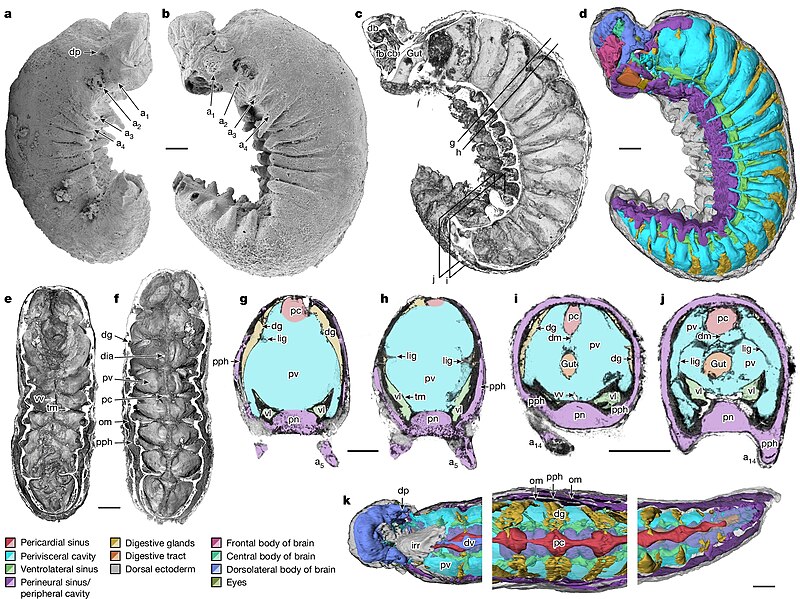
Description Anatomical overview of Youti yuanshi. YKLP 12387. a, External scanning electron microscopy, right side. Damage to posterior epidermis exposes lining of perivisceral cavity, demonstrating blind gut. b, External scanning electron microscopy, left side. c,g–j, Median virtual dissection from X-ray computed tomography (XCT) data (c), showing location of transverse slices intersecting digestive glands (g,i) and transverse membrane (h,j). d, Semi-manual segmentation of internal chambers from XCT data, viewed from the left side. Dorsolateral aspects of the peripheral cavity are omitted for clarity. e,f, Virtual dissection parallel to coronal plane, looking ventrally (e) and dorsally (f), showing digestive glands, pericardial sinus, transverse membranes within perivisceral cavity, and oblique membranes within peripheral cavity. g–j, XCT sections at positions indicated in c at position of digestive glands (g,i) and at position of ventrolateral lacunae and transverse membrane (h,j). g,h, Sections close to the anterior trunk, reflecting segments at late developmental stage. i,j, Sections close to the posterior trunk, showing superior preservation of internal tissue. k, Segmentation of internal chambers from XCT data, viewed from the dorsal perspective at anterior, middle and posterior trunk. Aspects of peripheral cavity are omitted for clarity. a, appendage; cb, central body of brain; db, dorsolateral body of brain; dia, diagenetic grain; dg, digestive gland; dm, dorsal membrane; dp, dorsal projection; dv, dorsal vessel; fb, frontal body of brain; irr, irregular chamber; lig, ligament; om, oblique membrane; pc, pericardial sinus; pph, peripheral cavity; pn, perineural sinus; pv, perivisceral cavity; tm, transverse membrane; vl, ventrolateral sinus; vv, ventral vessel. Scale bars=200 μm.
Date
Source Smith, Martin R.; Long, Emma J.; Dhungana, Alavya; Dobson, Katherine J.; Yang, Jie; Zhang, Xiguang (2024-07-31). "Organ systems of a Cambrian euarthropod larva". Nature. doi:10.1038/s41586-024-07756-8. ISSN 0028-0836.
Author Smith, Martin R.; Long, Emma J.; Dhungana, Alavya; Dobson, Katherine J.; Yang, Jie; Zhang, Xiguang
Permission
(Reusing this file)
w:en:Creative Commons
attribution
This file is licensed under the Creative Commons Attribution 4.0 International license.
You are free:
  • to share – to copy, distribute and transmit the work
  • to remix – to adapt the work
Under the following conditions:
  • attribution – You must give appropriate credit, provide a link to the license, and indicate if changes were made. You may do so in any reasonable manner, but not in any way that suggests the licensor endorses you or your use.

Captions

Add a one-line explanation of what this file represents

Items portrayed in this file

depicts

image/jpeg

File history

Click on a date/time to view the file as it appeared at that time.

Date/TimeThumbnailDimensionsUserComment
current01:59, 4 August 2024Thumbnail for version as of 01:59, 4 August 20242,126 × 1,591 (706 KB)SlvrHwkHigh-res version without full figure caption
03:44, 1 August 2024Thumbnail for version as of 03:44, 1 August 2024895 × 856 (581 KB)Shyamal{{Information |Description=Anatomical overview of Youti yuanshi |Source=Smith, Martin R.; Long, Emma J.; Dhungana, Alavya; Dobson, Katherine J.; Yang, Jie; Zhang, Xiguang (2024-07-31). "Organ systems of a Cambrian euarthropod larva". Nature. doi:10.1038/s41586-024-07756-8. ISSN 0028-0836. |Date=2024 |Author=Smith, Martin R.; Long, Emma J.; Dhungana, Alavya; Dobson, Katherine J.; Yang, Jie; Zhang, Xiguang |Permission={{cc-by-4.0}} |other_versions= }}

The following 2 pages use this file:

Global file usage

The following other wikis use this file:

Metadata