No higher resolution available.
Rms2.jpg (763 × 579 pixels, file size: 230 KB, MIME type: image/jpeg)
| This is a file from the Wikimedia Commons. Information from its description page there is shown below. Commons is a freely licensed media file repository. You can help. |
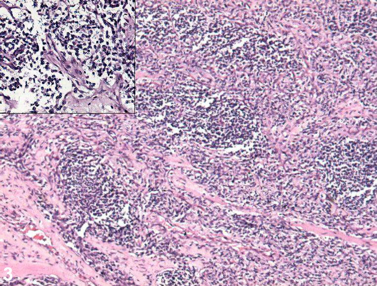
| Description | Photomicrograph showing nodules of tumor cells separated by hyalinised fibrous septae (50×, HE stain). Inset: Discohesive large tumor cells with hyperchromatic nucleus and scant cytoplasm (200×, HE stain). The diagnosis was postauricular congenital alveolar rhabdomyosarcoma |
| Date | |
| Source | Mahesha Vankalakunti, Ashim Das and Narasimhan KL Rao. Postauricular congenital alveolar rhabdomyosarcoma- a case report of an unusual entity. Diagnostic Pathology 2006, 1:37doi:10.1186/1746-1596-1-37 |
| Author | Mahesha Vankalakunti et al. |
This file is licensed under the Creative Commons Attribution 2.0 Generic license.
- You are free:
- to share – to copy, distribute and transmit the work
- to remix – to adapt the work
- Under the following conditions:
- attribution – You must give appropriate credit, provide a link to the license, and indicate if changes were made. You may do so in any reasonable manner, but not in any way that suggests the licensor endorses you or your use.
File history
Click on a date/time to view the file as it appeared at that time.
| Date/Time | Thumbnail | Dimensions | User | Comment | |
|---|---|---|---|---|---|
| current | 01:18, 16 January 2008 | | 763 × 579 (230 KB) | Filip em | {{Information |Description=Photomicrograph showing nodules of tumor cells separated by hyalinised fibrous septae (50×, HE stain). Inset: Discohesive large tumor cells with hyperchromatic nucleus and scant cytoplasm (200×, HE stain). The diagnosis was po |
File usage
The following page uses this file:
Global file usage
The following other wikis use this file:
- Usage on pl.wiki.x.io
- Usage on tr.wiki.x.io