File:Arvd MRI.jpg

Arvd_MRI.jpg (630 × 438 pixels, file size: 38 KB, MIME type: image/jpeg)

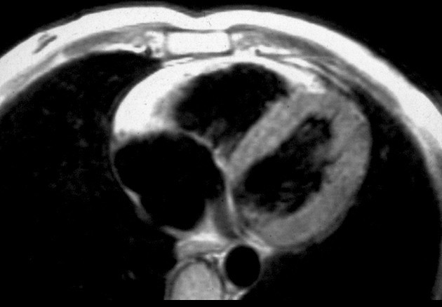
Description MRI in a patient affected by ARVC/D (long axis view of the right ventricle): note the transmural diffuse bright signal in the RV free wall on spin echo T1 (a) due to massive myocardial atrophy with fatty replacement (b).
Date 6 May 2008 (upload date)
Source Thiene et al. Orphanet Journal of Rare Diseases 2007 2:45 doi:10.1186/1750-1172-2-45
Author Thiene et al
Permission
(Reusing this file)
w:en:Creative Commons
attribution
This file is licensed under the Creative Commons Attribution 2.0 Generic license.
You are free:
  • to share – to copy, distribute and transmit the work
  • to remix – to adapt the work
Under the following conditions:
  • attribution – You must give appropriate credit, provide a link to the license, and indicate if changes were made. You may do so in any reasonable manner, but not in any way that suggests the licensor endorses you or your use.

Captions

Add a one-line explanation of what this file represents

Items portrayed in this file

depicts

image/jpeg

85087208af436dded8e501c0167d06a9dd9d10db

38,445 byte

438 pixel

630 pixel

File history

Click on a date/time to view the file as it appeared at that time.

Date/TimeThumbnailDimensionsUserComment
current15:22, 6 May 2008Thumbnail for version as of 15:22, 6 May 2008630 × 438 (38 KB)Filip em{{Information |Description=MRI in a patient affected by ARVC/D (long axis view of the right ventricle): note the transmural diffuse bright signal in the RV free wall on spin echo T1 (a) due to massive myocardial atrophy with fatty replacement (b). |Source

The following page uses this file:

Global file usage

Metadata